Scientific instrumentation is an important part of our laboratory, particularly for biophysics activities. The in-house design of the vast majority of our measuring instruments has been the originality and strength of our laboratory since its creation. This approach began in the 1960s under the leadership of Pierre Joliot with the creation of a modulated oxygen electrode, which allowed him to resolve the S states of photosynthesis. It continued throughout his career with the rapid recruitment of Daniel Béal, a research engineer to support him. This collaboration led to the development of numerous innovative optical difference absorption spectroscopy devices and resulted in the creation of a patent and the commercialization of a measuring device called JTS (Joliot Type Spectrometer) by the company Biologic. Currently, the unit’s scientific instrumentation is managed by Julien Sellés, a CNRS research engineer. The development of new instruments as well as the improvement of existing devices is done in close collaboration with Benjamin Bailleul (CRCN), Wojciech Nawrocki (CRCN) and Christopher Larran (IR CDD CNRS). Collaborations with private actors are also established with JBeamBio, a scientific instrumentation company founded by Daniel Béal, ABMP, a company specialized in electronic design, and API whose core business is mechanical design.
This approach of “instrumentation adapted to researchers” rather than the reverse, has allowed the laboratory throughout its existence to be a pioneer in certain research fields related to photosynthesis. It has also enabled the establishment of numerous lasting international collaborations with prestigious research institutes.
From a practical point of view, the laboratory currently has the following instruments:
Time-Resolved Difference Absorption Spectrophotometer or JTS:
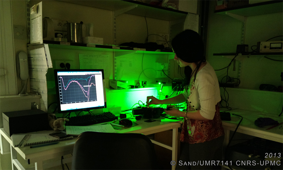
This type of spectrophotometer developed in the laboratory is based on the principle of pump-probe spectroscopy [1, 2]. A high-intensity light beam, called a pump, is sent to the sample to trigger photosynthesis. A second beam of low intensity and very short duration, called a probe, is then sent and allows probing the difference in absorption between a sample where photosynthesis is active and a sample in darkness. The measured absorption difference then allows us to trace back to the oxidation-reduction state of the various actors in photosynthesis, such as photosystem I, cytochrome b6f or plastocyanins, but also to the activity of ATP-synthase. This technique allows working both in vivo on whole cells or leaves and in vitro. The following configurations are available:
- 2 “LED” JTS: excitation is done either by a nanosecond laser flash (YAG amplitude laser) at 695 nm or 532 nm. The probe pulse (20 μs) is obtained using a white LED filtered by interference filters. These devices have a temporal resolution of 150 μs and allow absorption measurements at specific wavelengths (520, 540, 554, 564, 573, 580, 700, 715, 730 nm).
- 1 “monochromator” JTS: identical to the LED JTS, the spectral selection of the probe beam is done through a monochromator, which allows a continuous measurement between 500 nm and 600 nm.
- 1 “OPO” JTS: Excitation is done by a nanosecond laser flash (OPO pumped by a YAG) and can vary continuously from 400 nm to 800 nm. The probe pulse comes from a broadband OPO laser (5ns). This device has a temporal resolution of 10 ns and a spectral resolution ranging from 260 nm to 1500 nm.
Fluorescence Spectrophotometer:
The analysis of fluorescence emitted by chlorophylls is a powerful and widely used tool to study in particular the photochemical efficiency of the photosynthetic chain. The light energy absorbed by a photosynthetic organism can be used according to 3 processes: i) photochemistry to start the photosynthesis process ii) dissipation in the form of heat iii) dissipation in the form of fluorescence. Since these three processes are in competition, the measured fluorescence gives us an indirect relative measurement of the efficiency of photosynthesis and in particular of photosystem II. The available equipment is as follows:
- A fluorometer called “liquid Fluo“: allows measuring the amount of fluorescence emitted, only on a liquid sample. All the collected fluorescence is spectrally integrated by a photodiode.
- A fluorometer called “77 K“: allows spectrally resolved fluorescence measurements on liquid samples. This equipment allows working at room temperature or at liquid nitrogen temperature.

- A fluorometer called “SpeedZen“: allows measuring fluorescence with spatial resolution. This type of setup is particularly suitable for measurements on leaves or multi-well plates. This technique is a powerful screening tool for studies where the photosynthetic chain is disturbed, due to the large number of possible experimental conditions simultaneously [3].
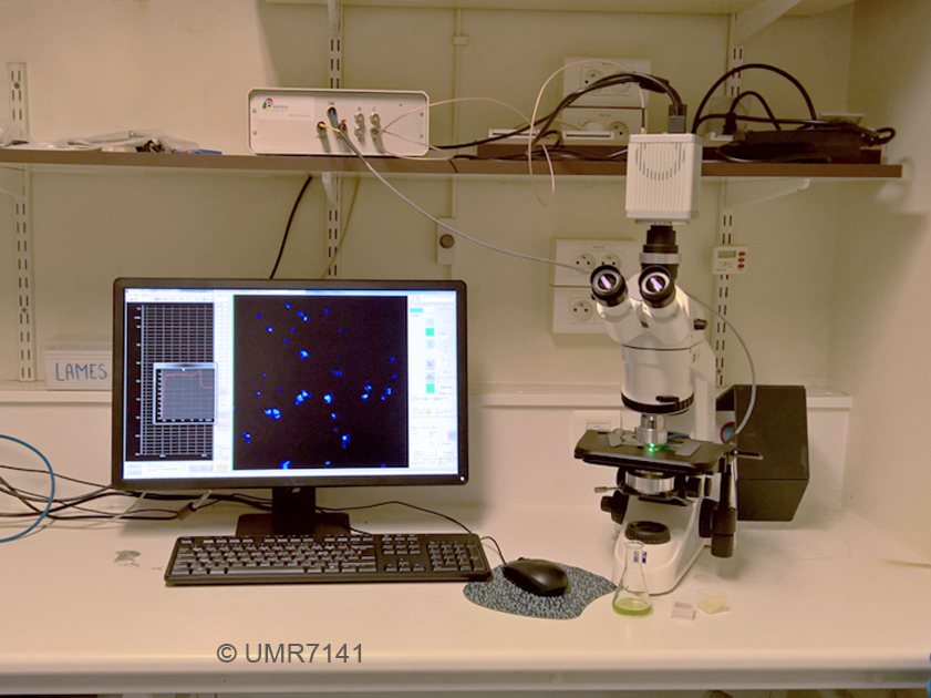
- A fluorometer called “micro-SpeedZen“: identical to the SpeedZen but mounted on a microscope stand to allow measurements on a single cell.
- A time-resolved fluorimeter (Time-Correlated Single Photon Counting type) : allows measurements of the excited state lifetimes across the emission spectrum with a ~50 ps time resolution.
Modulated Oxygen Electrode:
The laboratory has recently acquired an oxygen electrode similar to that developed by Pierre Joliot in the 1960s. This type of device consists of a platinum electrode and a silver/silver chloride electrode between which a negative voltage of 0.75 V is applied. A uniform layer of unicellular algae can be uniformly deposited on the platinum electrode, and the emission of oxygen causes an oxidation-reduction reaction, which generates a measurable current. The particularity of our electrode is the illumination of the algae, which is modulated sinusoidally and allows measuring the average rate of oxygen emission [4].
Biolistic Transformation Device:
![]()
To transform the chloroplast genome of Chlamydomonas reinhardtii, we use a particle gun (biolistic transformation method) built in the laboratory and adapted from a similar device built at the University of Geneva. A jet of pressurized helium (pressure 7 bars, jet duration 20 ms) allows projecting tungsten particles (Ø 1μm) coated with DNA (2 μg of supercoiled plasmid per shot) onto a cellular carpet placed in a vacuum environment (approximately 1-2 107 cells; vacuum 48 mbars; length of the bead path 15 cm). We obtain up to 1000 transformant clones per shot.
[1] Joliot P., Béal D. and Friley B. (1980) A new spectrophotometric method for the study of photosynthetic reactions. Journal de Chimie Physique et de Physico-Chimie Biologique, 77(3) :209-216
[2] Béal D., Rappaport F. and Joliot P. (1999) A new high-sensitivity 10-ns time-resolution spectrophotometric technique adapted to in vivo analysis of the photosynthetic apparatus. Review of scientific instruments 70(1) : 202-207
[3] Johnson X, Vandystadt G, Bujaldon S, Wollman F-A, Dubois R, Roussel P, Alric J and Béal D (2009) A new setup for in vivo fluorescence imaging of photosynthetic activity. Photosynth Res, 102(1):85-93
[4] Joliot Pierre (1956) Dispositif ampérométrique de mesure de la photosynthèse. C.R. Acad. Sci. Paris, 243(7):677-80